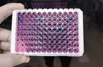
When you rely on 2D Gel Electrophoresis, your results are only as valuable as the quality of the data you generate and interpret. Whether you are studying disease biomarkers, comparing protein expression, validating research findings, or supporting product development, every gel contains valuable information waiting to be uncovered. By optimizing each stage of your workflow, you can improve reproducibility, identify more proteins, and gain deeper biological insights.
Instead of focusing solely on producing clear gel images, you should aim to maximize every piece of information your experiment provides. From sample preparation to image analysis, each decision influences the quality and usefulness of your final dataset.
Why Data Quality Matters in 2D Gel Electrophoresis
Two-dimensional gel electrophoresis separates proteins based on two independent characteristics: isoelectric point (pI) and molecular weight. This powerful combination allows you to visualize hundreds or even thousands of proteins simultaneously.
However, obtaining high-quality data requires more than running a successful gel. Poor sample preparation, inconsistent experimental conditions, or inadequate analysis can reduce spot resolution, introduce artifacts, and limit meaningful interpretation.
When every step is optimized, you can:
- Detect low-abundance proteins
- Improve spot resolution
- Increase experimental reproducibility
- Identify subtle protein expression changes
- Generate stronger evidence for publication or regulatory submissions
Start with High-Quality Sample Preparation
The quality of your sample determines the quality of your results.
Before beginning your experiment, ensure your samples are:
- Fresh and properly stored
- Free from contaminants
- Compatible with electrophoresis buffers
- Properly quantified for protein concentration
Removing salts, lipids, nucleic acids, and other interfering substances significantly improves protein separation.
Consistent sample handling also reduces variability between experimental runs.
Choose the Right Protein Extraction Method
Different sample types require different extraction strategies.
Consider factors such as:
- Tissue type
- Cell line
- Biological fluid
- Membrane proteins
- Protein solubility
Using optimized lysis buffers helps preserve proteins while minimizing degradation.
Protease and phosphatase inhibitors should be included whenever appropriate to protect sensitive proteins throughout extraction.
Optimize the First-Dimension Separation
Isoelectric focusing is one of the most critical stages of the experiment.
To improve separation:
- Select an appropriate pH range
- Avoid overloading protein samples
- Ensure complete sample solubilization
- Follow recommended voltage programs
- Maintain consistent running conditions
Proper focusing allows proteins with similar molecular weights but different isoelectric points to separate more effectively.
Improve SDS-PAGE Performance
The second dimension separates proteins by molecular weight.
For better results:
- Use freshly prepared gels
- Maintain consistent gel composition
- Standardize electrophoresis conditions
- Avoid overheating during runs
- Use appropriate molecular weight markers
Even small inconsistencies can affect migration patterns and reduce data accuracy.
Prevent Common Experimental Errors
Many data quality issues originate from preventable mistakes.
Watch for:
- Protein degradation
- Incomplete focusing
- Air bubbles
- Gel tearing
- Uneven staining
- Sample contamination
- Inconsistent loading volumes
Developing standardized laboratory protocols minimizes these variables and improves reproducibility across multiple experiments.
Use Sensitive Protein Staining Methods
Protein visualization directly affects how much information you can collect.
Depending on your research goals, choose staining methods that provide:
- High sensitivity
- Wide dynamic range
- Low background noise
- Excellent reproducibility
- Compatibility with downstream protein identification
Consistent staining improves spot detection and quantitative analysis.
Capture High-Resolution Images
Image quality directly influences data analysis.
For accurate spot detection:
- Use calibrated imaging systems
- Avoid image saturation
- Maintain identical imaging settings across experiments
- Capture images at maximum practical resolution
Well-documented imaging procedures also improve long-term project consistency.
Analyze Images with Reliable Software
Image analysis transforms gel images into meaningful biological information.
Modern software can help you:
- Detect protein spots automatically
- Compare multiple gels
- Quantify protein abundance
- Normalize datasets
- Reduce operator bias
- Generate statistical reports
Always verify automated spot matching manually to ensure accuracy.
Validate Significant Protein Changes
Not every visible difference represents a biologically meaningful change.
After identifying proteins with altered expression, validate findings using complementary techniques whenever possible.
Validation strengthens confidence in your conclusions and supports publication-quality research.
Standardize Every Experiment
Reproducibility remains one of the biggest challenges in proteomics.
Create standardized procedures for:
- Sample collection
- Storage conditions
- Protein extraction
- Electrophoresis
- Staining
- Imaging
- Data processing
Documenting every experimental parameter makes future comparisons far more reliable.
Collaborate with Experienced Protein Analysis Specialists
Working with experienced laboratories can significantly improve data quality.
Professional laboratories often provide:
- Optimized protocols
- Advanced instrumentation
- Experienced technical staff
- High-resolution imaging
- Comprehensive data analysis
- Quality assurance procedures
If your project requires dependable protein separation and comprehensive analysis, explore advanced 2D Gel Electrophoresis protein analysis services for complex proteomics research to improve experimental outcomes.
You can also learn more about the extensive protein analysis capabilities offered by Kendrick Labs, Inc. for specialized electrophoresis and proteomics solutions.
Best Practices for Maximizing Your Data
To consistently obtain high-value datasets:
- Use clean, high-quality samples.
- Standardize every laboratory procedure.
- Optimize both dimensions of separation.
- Select sensitive staining techniques.
- Capture high-resolution digital images.
- Perform careful image analysis.
- Validate significant protein changes.
- Maintain complete experimental documentation.
- Repeat experiments with biological replicates.
- Work with experienced proteomics specialists when needed.
Following these practices helps you generate more reliable, reproducible, and publication-ready results.
Conclusion
Maximizing data from 2D Gel Electrophoresis requires attention to every stage of the experimental workflow. High-quality samples, optimized separation conditions, careful staining, accurate imaging, and robust data analysis all contribute to more meaningful protein identification and quantification.
When you combine standardized laboratory practices with experienced analytical support, you improve confidence in every result while uncovering deeper biological insights. Whether your goal is biomarker discovery, protein characterization, or comparative proteomics, investing in quality throughout the workflow produces data you can trust.
For expert guidance on your next proteomics project, Contact us today to discuss your protein analysis requirements with experienced specialists.
Frequently Asked Questions
What is 2D Gel Electrophoresis used for?
2D Gel Electrophoresis is used to separate complex protein mixtures based on isoelectric point and molecular weight, enabling researchers to compare protein expression, detect biomarkers, and study protein modifications.
How can you improve protein spot resolution?
You can improve spot resolution by using high-quality samples, optimizing protein extraction, selecting the correct pH range, preventing contamination, and maintaining consistent electrophoresis conditions.
Why is sample preparation so important?
Proper sample preparation removes contaminants, preserves protein integrity, and improves protein separation, resulting in more reliable and reproducible experimental data.
What factors reduce data quality in 2D Gel Electrophoresis?
Common factors include protein degradation, inconsistent sample loading, poor staining, incomplete focusing, contaminated samples, imaging errors, and inadequate data analysis.
Why should researchers use experienced protein analysis laboratories?
Experienced laboratories provide optimized protocols, advanced instrumentation, quality-controlled workflows, expert analysis, and reproducible results that improve confidence in proteomics research.